
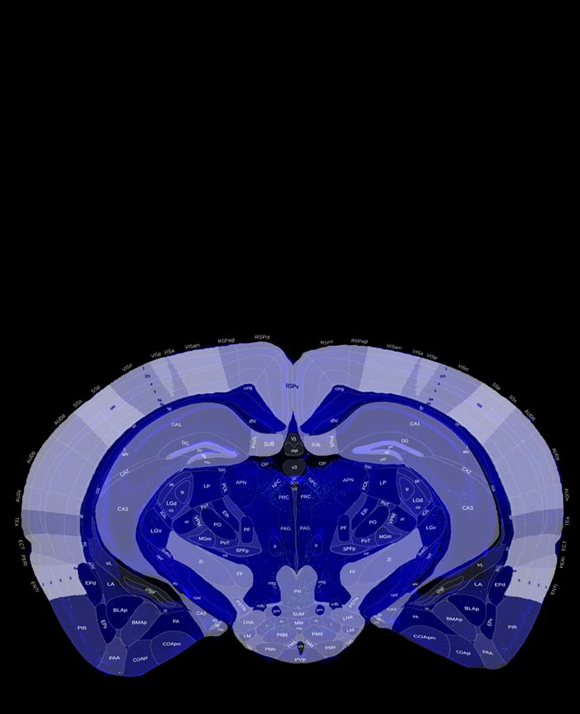
One-click Atlas Registration
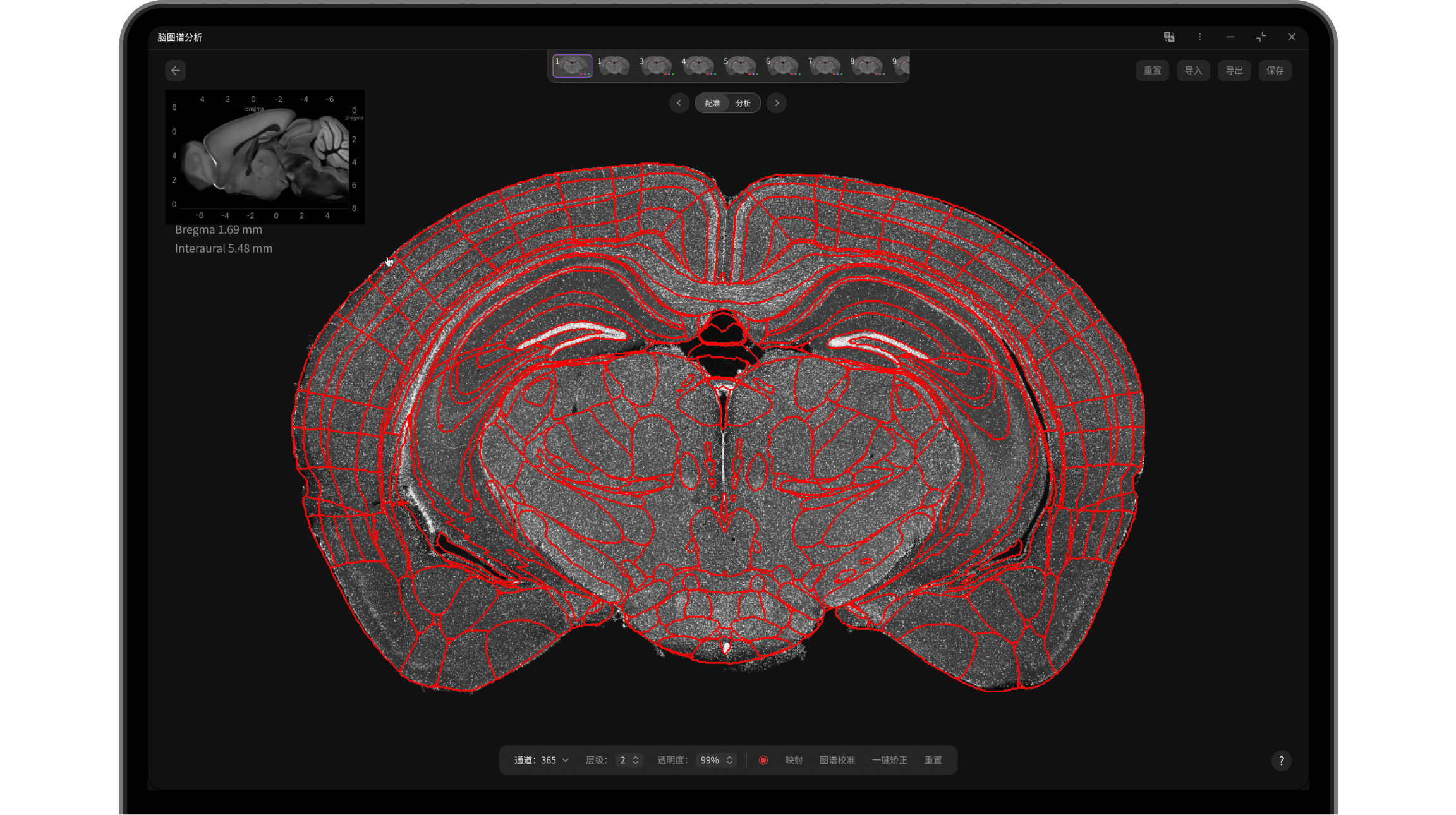
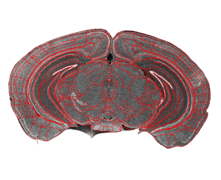
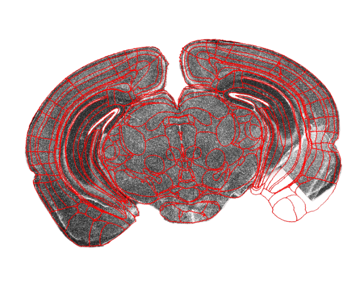
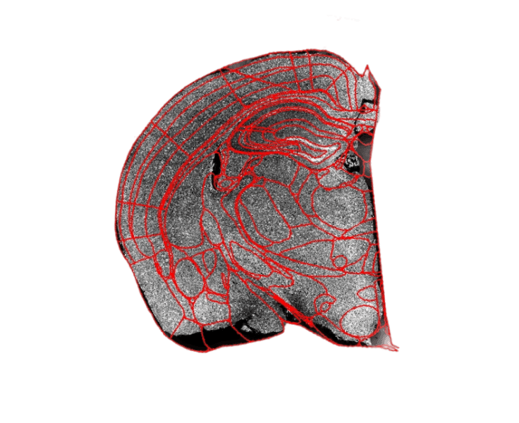
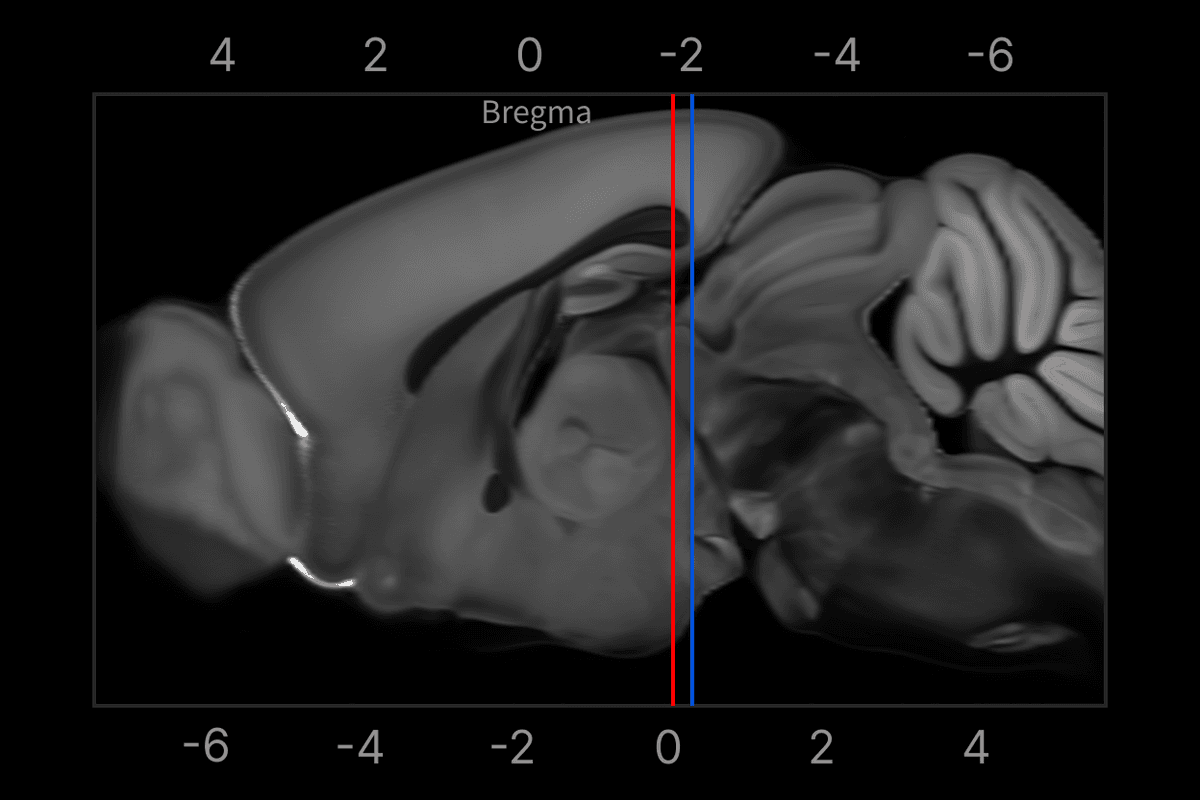
One-click registration
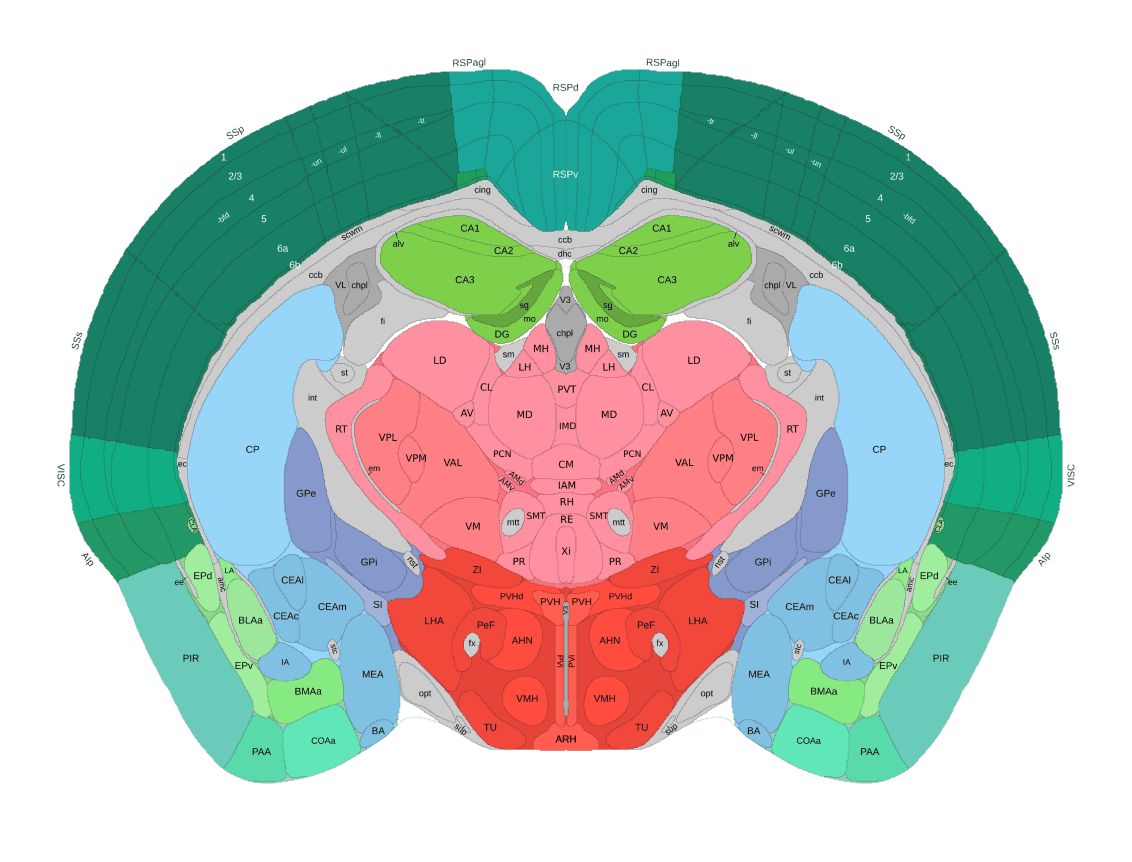
Automatically identifies brain slices and matches them to the Allen Brain Atlas
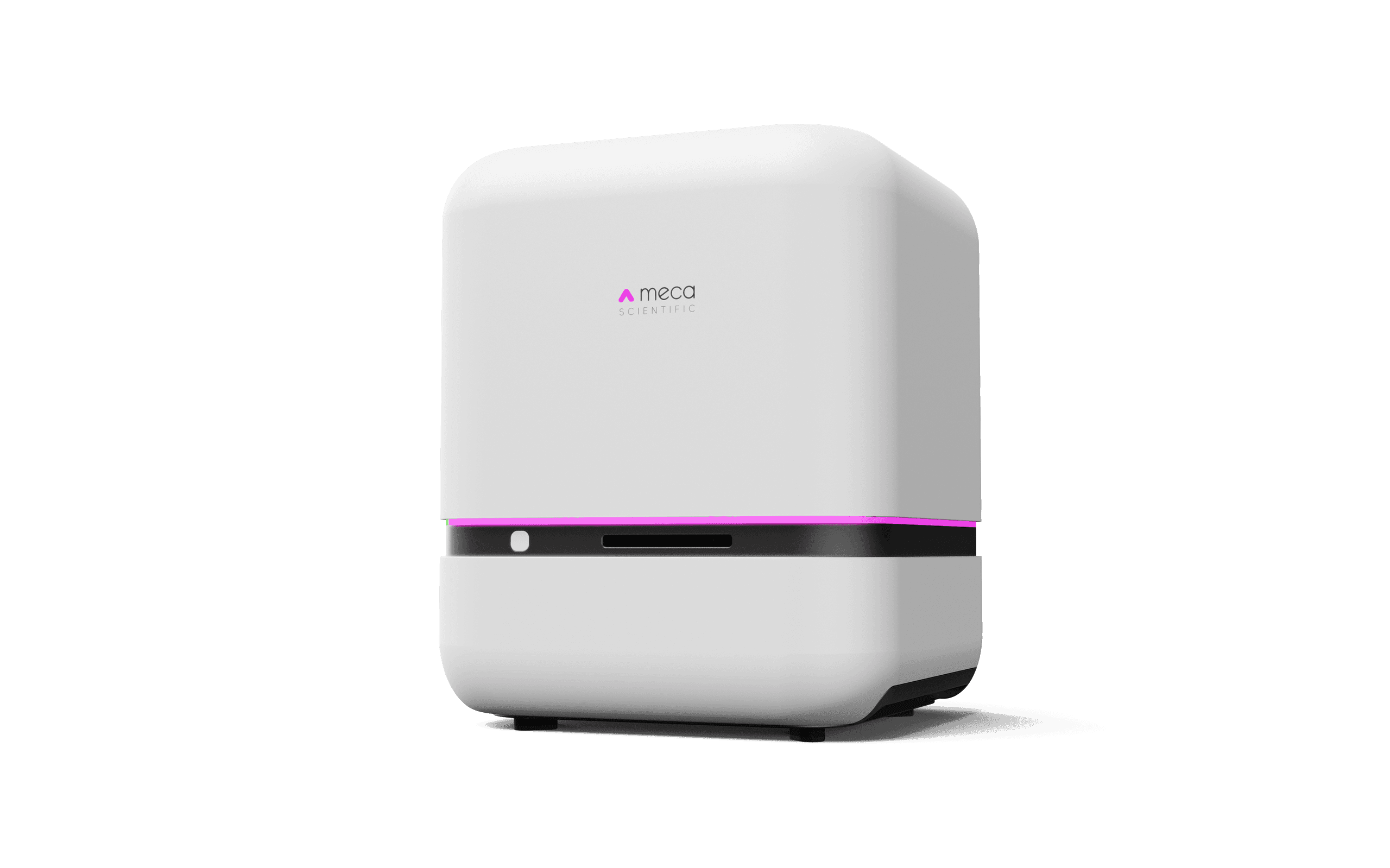
PanoBrain / Fully Automatic Brain Slice Analyzer
PanoBrain rapidly scans brain slices to capture high-resolution digital panoramic images, then automatically performs quantitative analysis such as brain-region segmentation, atlas registration, and cell counting. It automates the full workflow from imaging to data processing and reshapes neuroscience research workflows
Apply for TrialOne-click Atlas Registration
Automatically identifies brain slices and matches them to the Allen Brain Atlas
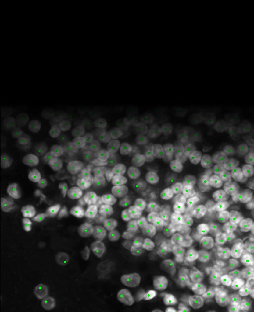
Cell Recognition and Counting
Reduces manual intervention while ensuring data consistency and accuracy, facilitating the extraction of reliable experimental results
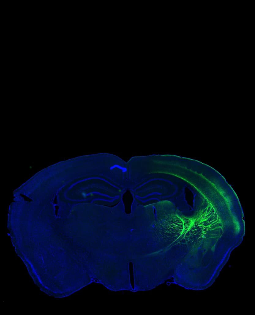
Intelligent Scan Path Planning
Automatically identifies ROIs and plans scan paths. A brain slice scan takes less than 1 minute; with a 10X objective and DAPI & GFP dual channels, a single brain slice takes only 40 seconds
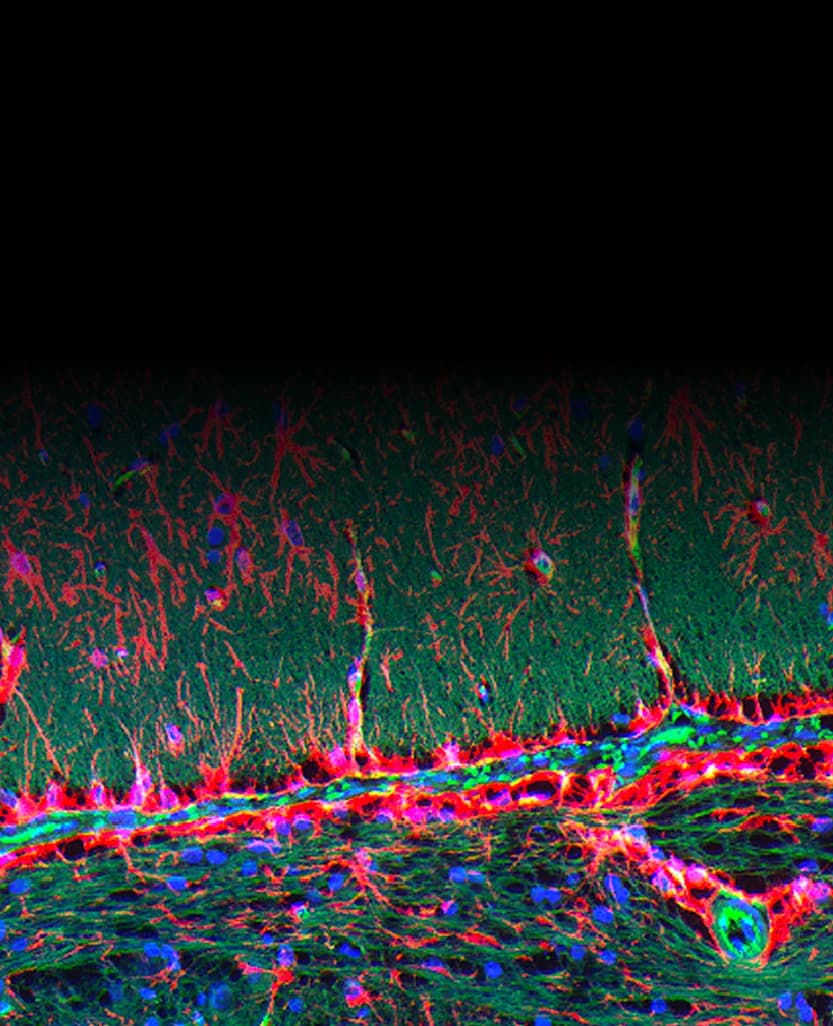
High-precision Autofocus
Precisely focuses on the surface of thick brain slices... and keeps focal planes consistent across all channels, generating high-quality multi-channel data
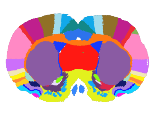

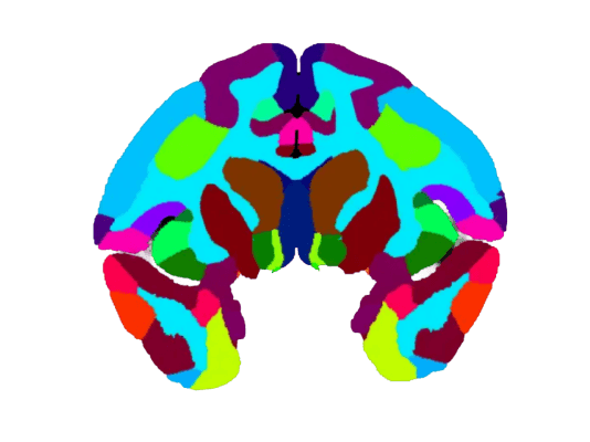

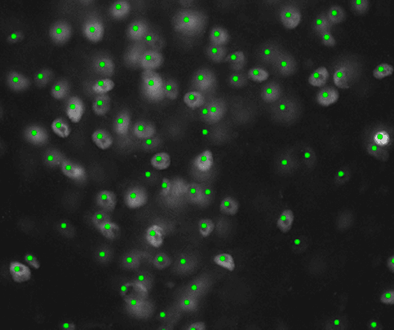
Supports automatic recognition of DAPI, c-Fos, neurons, glial cells, and other signals, with statistics for positive signal count, area, and fluorescence intensity
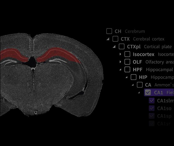
Supports whole-brain, left-right brain, and custom brain-region statistics to meet diverse counting needs
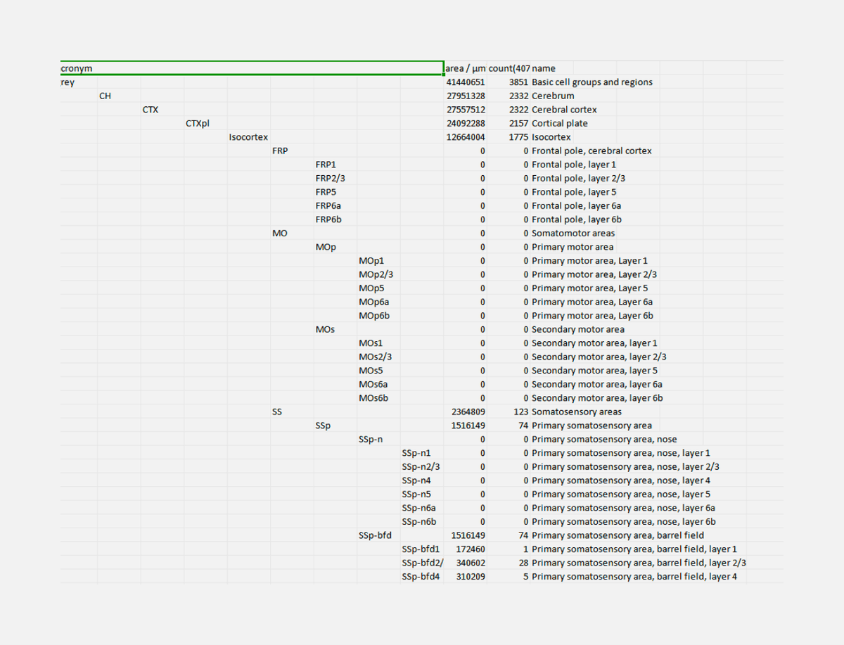
Exports results to Excel in a hierarchical format, clearly linking parent and child brain-region data, making complex anatomical statistics easy to interpret
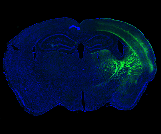
Automatically identifies ROIs and plans scan paths, keeping each brain slice scan under 1 minute
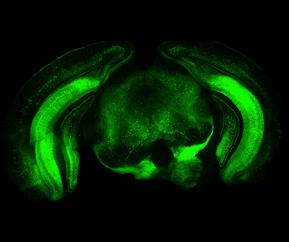
Uniform illumination coupled with global background correction achieves up to 99% field uniformity, effortlessly yielding high-quality multi-channel fluorescence images
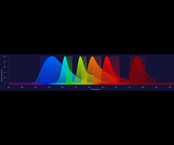
Universal fluorescence channel configurations cover common fluorescence imaging applications, with customizable narrow-band excitation and emission filters to eliminate crosstalk anxiety
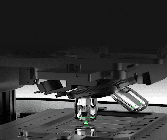
Strobe illumination provides millisecond-level high instantaneous power only during camera exposure, reducing phototoxic effects caused by continuous illumination
Side-mounted scattered LED illumination enables high-contrast visualization of fluorescent samples, eliminating the need for manual marker-pen positioning
An embedded ring light shows scan status and progress through color and completion state, making experiment progress easy to track
Scan parameters, image contrast, analysis regions, and export size settings can be saved as templates for fast reuse
Supports batch export of multi-channel fluorescence images as single-channel pseudo-color or grayscale, and multi-channel overlays. It seamlessly combines flexible image type selection with highly efficient batch processing
Move brain slice scanning, atlas registration, and quantitative analysis into a more efficient automated workflow
Apply for Trial